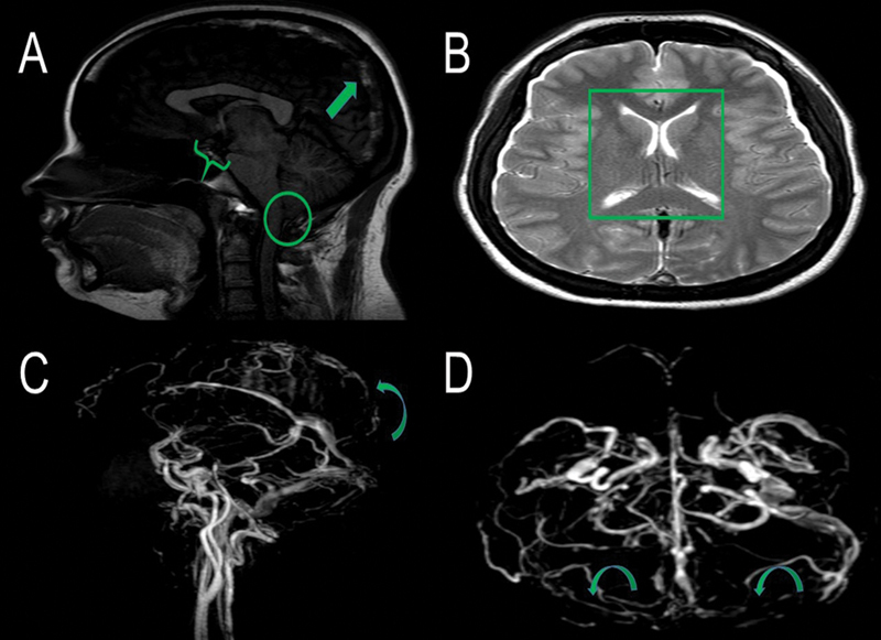
Figure 1.

Brain magnetic resonance imaging (MRI) sagittal T1-weighted image (without contrast administration) (A) shows a hyperintense thrombus in the superior sagittal sinus (arrow), decreased mamillopontine distance (0,3 cm) (brace) and cerebelar tonsilar ectopia (0.5 cm) (CIRCLE). Brain MRI axial T2 weighted (B) image demonstrates collapsed lateral ventricles space (square). Brain MRI venography (C and D) shows a contrast filling defect in the topography of superior sagittal and bilateral transverse sinus suggesting CVT (curve arrows)
