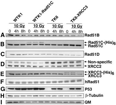
Figure 2.
Constitutive and X-ray-induced levels of the Rad51 paralogs in different human cell lines. Western blot analysis using different antibodies was used to determine the constitutive level of the Rad51 paralogs and of hRad51, and the level of these proteins 4 and 8 h after treatment with 10 Gy X-rays. P53 was used as a control for the X-ray treatment (see text), and either β-tubulin or transcription factor QM was used as loading standards for each western blot (only one representative blot for each is shown). Unlike Figure 1, the detection of XRCC3 here was done using an antibody from Novus that only weakly recognizes the recombinant XRCC3-(His)6 protein (see text).

