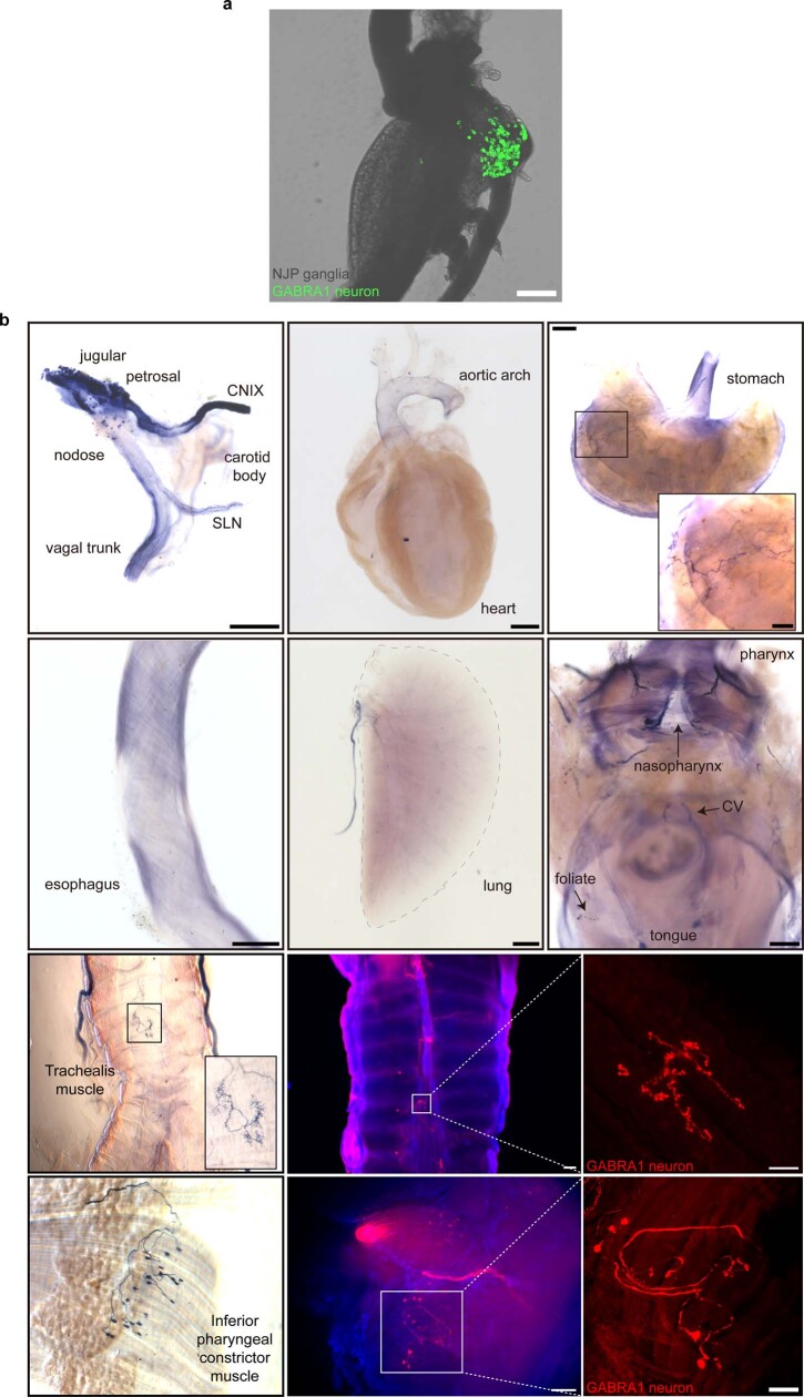
Extended Data Fig. 10. Sparse innervation of internal organs by GABRA1 NJP neurons.
a, Native GFP fluorescent signals in wholemount preparations of NJP ganglia from Gabra1-ires-cre; lsl-L10 GFP, scale bar: 200 μm. b, NJP ganglia of Gabra1-ires-Cre mice were injected bilaterally with Cre-dependent AAV-flex-AP or AAV-flex-tdTomato and axons were visualized in fixed wholemount tissue preparations using either a colorimetric alkaline phosphatase substrate (top two rows, left images in bottom two rows) or tdTomato immunostaining (right two images in bottom two rows). Scale bars (left to right) top row: 500, 1000, 1000 (inset: 250) μm; 2nd row: 500, 1000, 500 μm; 3rd row: 200, 50 μm; bottom row: 200, 100 μm. Images are representative of three independent experiments involving GFP and tdTomato and two independent experiments involving alkaline phosphatase.

