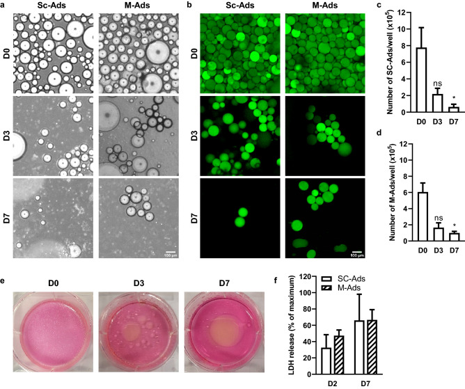
Figure 1.
Like SC-Ads, M-Ads rapidly die when cultured in 2D. (a) Representative phase-contrast images taken under light microscope of human SC-Ads and M-Ads cultured in 2D for the indicated times; Scale bar, 100 µm. (b) Representative images of BODIPY® 493/503 (neutral lipids, green) stained primary SC-Ads and M-Ads in 2D culture for the indicated times; Scale bar, 100 µm. (c,d) Number of SC-Ads (c) and M-Ads (d) cultured in 2D over time (n = 3). (e) Representative pictures of the culture wells of M-Ads grown in 2D at indicated times points. (f) Quantification of LDH release in medium during 2D culture at D2 and D7 for SC-Ads and M-Ads (n = 3). The results are expressed as the percentage of the value obtained when the whole cell population (same cell number than D0) is lysed. The histograms represent mean ± SEM, ns non-significant, *P < 0.05.

