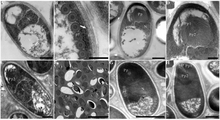
Figure 5.
Electron microscopy of Pseudokabatana alburnus isolated from different fish. (A) A mature spore of P. alburnus isolated from Cultrichthys erythropterus showing six coils of polar filament (pf), a vacuole (V) and a large nucleus (N). Scale bar = 500 nm. (B) Magnification of trilaminar spores wall showing an electron-dense exospore (Ex), an electron-translucent endospore (En) and a plasma membrane (arrow). Scale bar = 200 nm. (C) A mature spore of P. alburnus isolated from C. erythropterus with bipartite polaroplast, a vacuole and a large nucleus. Scale bar = 500 nm. (D) Magnification of bipartite polaroplast (Pp1, Pp2). Scale bar = 200 nm. (E) A mature spore of P. alburnus isolated from Pseudolaubuca engraulis with a bipartite polaroplast (Pp1, Pp2), a large nucleus (N) and the isofilar polar filaments (pf). Scale bar = 500 nm. (F) Mature spores occur in direct contact with the host cell cytoplasm. Scale bar = 5 μm. (G) A mature spore of P. alburnus isolated from Elopichthys bambusa showing 5–6 coils of polar filament (pf), a vacuole (V) and a large nucleus (N). Scale bar = 500 nm. (H) A mature spore of P. alburnus isolated from E. bambusa showing bipartite polaroplast (Pp1, Pp2) and a mushroom-shaped anchoring disk (AD). Scale bar = 500 nm.

