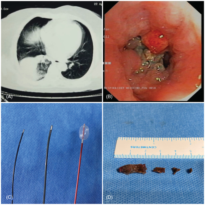
FIGURE 1.

(A) CT chest showing mass like consolidation in the right lower lobe, (B) endobronchial view showing impacted foreign body material with granulation tissue in RB10, (C) 1.1 mm, 1.7 mm Erbe cryoprobe and 4 Fr Fogarty balloon, and (D) retrieved clove
