
FIGURE 2.
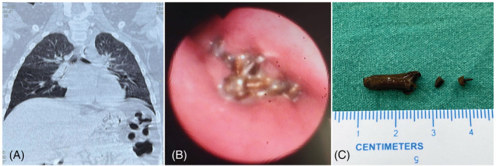
(A) Coronal plane CT showing obstruction in the right intermediate bronchus, (B) endobronchial view at the level of right main bronchus showing impacted clove with granulation tissue in the proximal right intermediate bronchus, and (C) retrieved clove