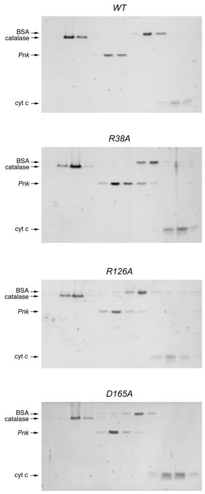
Figure 3.
Glycerol gradient sedimentation of Pnk and Pnk-Ala mutants. Sedimentation analysis was performed as described in Materials and Methods. The distributions of Pnk (either wild-type, R38A, R126A or D165A as indicated) and the marker proteins catalase, BSA and cytochrome c in each gradient were analyzed by SDS–PAGE. Scans of the Coomassie blue stained gels are shown.

