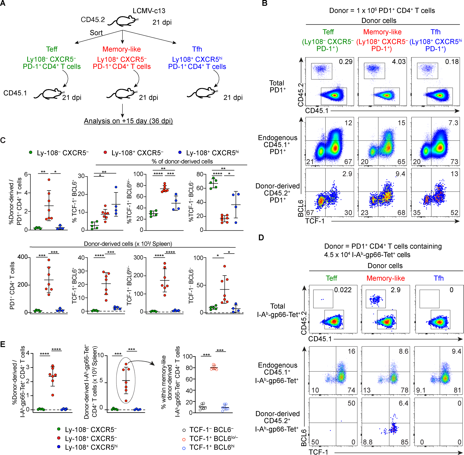
Figure 4. TCF-1+ BCL6lo/− PD-1+ CD4+ T cells are capable of superior expansion and differentiation into both Teff and Tfh cells following adoptive transfer. (See also Figure S5).

(A) Experimental design of adoptive transfer of distinct PD-1+ CD4+ T cell populations from LCMV-c13 infected mice to infection-matched recipient mice for the experiments shown in (B-E). For the experiments shown in (B) and (C), 1 × 10⁶ PD-1+ CD4+ T cells were transferred. For the experiments shown in (D) and (E), 1–2 × 10⁶ PD-1+ CD4+ T cells containing 4.5 × 10⁴ Tet+ cells were transferred.
(B-E) Representative flow cytometry plots showing frequencies of total donor-derived cells (B) and Tet+ donor-derived cells (D) and their expression of TCF-1 and BCL6 15 days after transfer. Pooled data from 2 experiments with n = 2–4 / group / experiment are shown in (C, E) with mean±SD and statistical analysis by One-way ANOVA. Frequencies and total numbers of donor-derived cells in (B, C) were normalized to 1 × 10⁶ transferred donor cells, and frequencies and total numbers of Tet+ donor-derived cells in (D, E) were normalized to 4.5 × 10⁴ Tet+ transferred donor cells.
