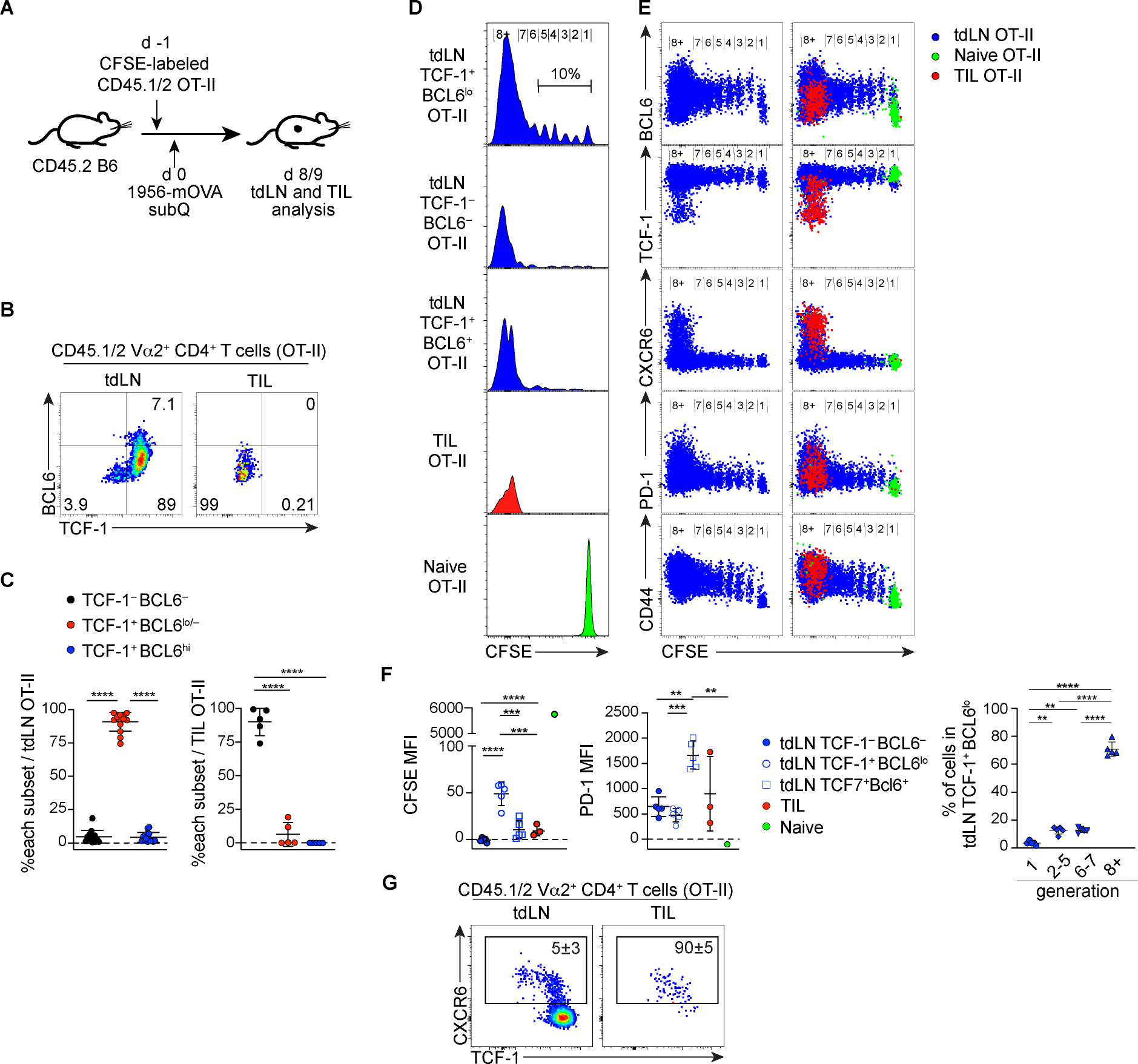
Figure 7. Antigen-specific CD4+ T cells differentiate into TCF-1+ PD-1+ cells following cell division in tumor-draining lymph nodes, but not in the tumor microenvironments.

(A) Experiment design to analyze CD4+ T cell response to tumor antigen.
(B-C) Representative plots showing expression of TCF-1 and BCL6 by donor-derived OT-II CD4+ T cells harvested from tumor (TIL) and the tumor draining lymph node (tdLN). Data pooled from two experiments (n = 6 – 8 / experiment) and shown with mean±SD.
(D-F) CFSE dilution and expression of BCL6, TCF-1, CXCR6, PD-1 and CD44 by donor-derived OT-II cells. The CFSE level of naive OT-II cells was determined by recipient mice without tumor transplantation sacrificed at the same time points. Left panels in (E) show tdLN-derived OT-II cells without overlay of TIL or naive cells. Pooled data are shown with mean±SD in (F) with assessment of statistical differences by one-way ANOVA.
(G) Representative plots showing expression of CXCR6, TCF-1 and BCL6 by donor-derived OT-II CD4+ T cells harvested from tdLN and the tumor. Numbers show mean±SD.
