
Fig. 1 |. The MIPSA method.
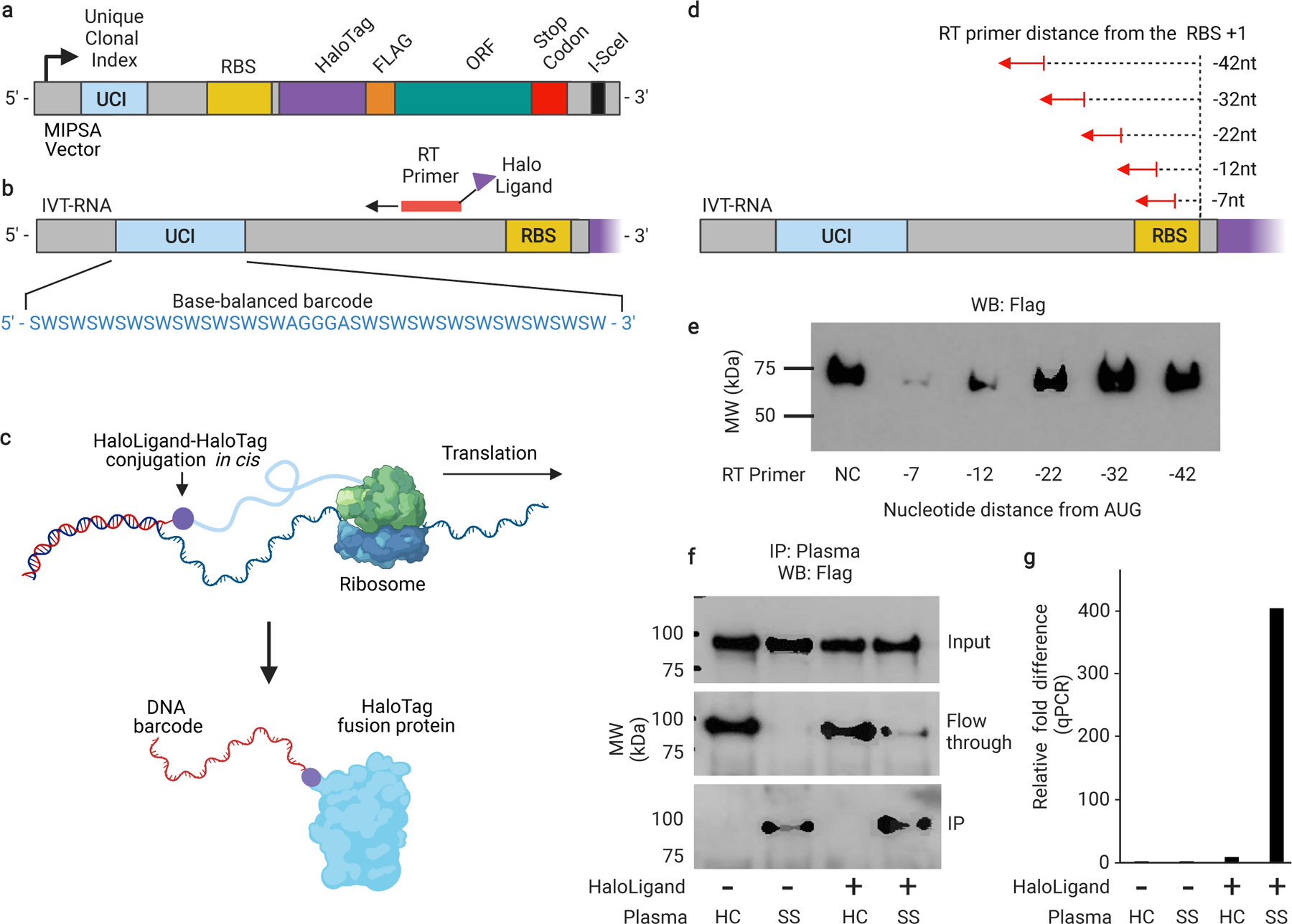
a, Schematic of the recombined pDEST-MIPSA vector with key components highlighted: unique clonal identifier (UCI, blue), ribosome binding site (RBS, yellow), N-terminal HaloTag (purple), FLAG epitope (orange), open reading frame (ORF, green), and the I-SceI restriction endonuclease site (black) for vector linearization. b, Schematic showing in vitro transcribed (IVT) RNA from the vector template shown in (a). Isothermal base-balanced UCI sequence: (SW)18-AGGGA-(SW)18. c, Cell-free translation of the RNA-cDNA shown in (b). HaloTag protein forms a covalent bond with the HaloLigand-conjugated UCI-containing cDNA in cis during translation. d, RT primer positions tested for impact on translation. e, α-FLAG western blot analysis of translation in presence of RT primers depicted in (d) (NC, negative control, no RT primer). f, Western blot analysis of TRIM21 protein translated from RNA carrying the UCI-cDNA primed from the −32 position, either conjugated (+) or not (−) with the HaloLigand. Sjogren’s Syndrome, SS; Healthy Control, HC. g, qPCR analysis of the IPed TRIM21 UCI. Fold-difference is by comparison with the HaloLigand (−) HC IP (technical replicates, n = 2).