
Figure 1.
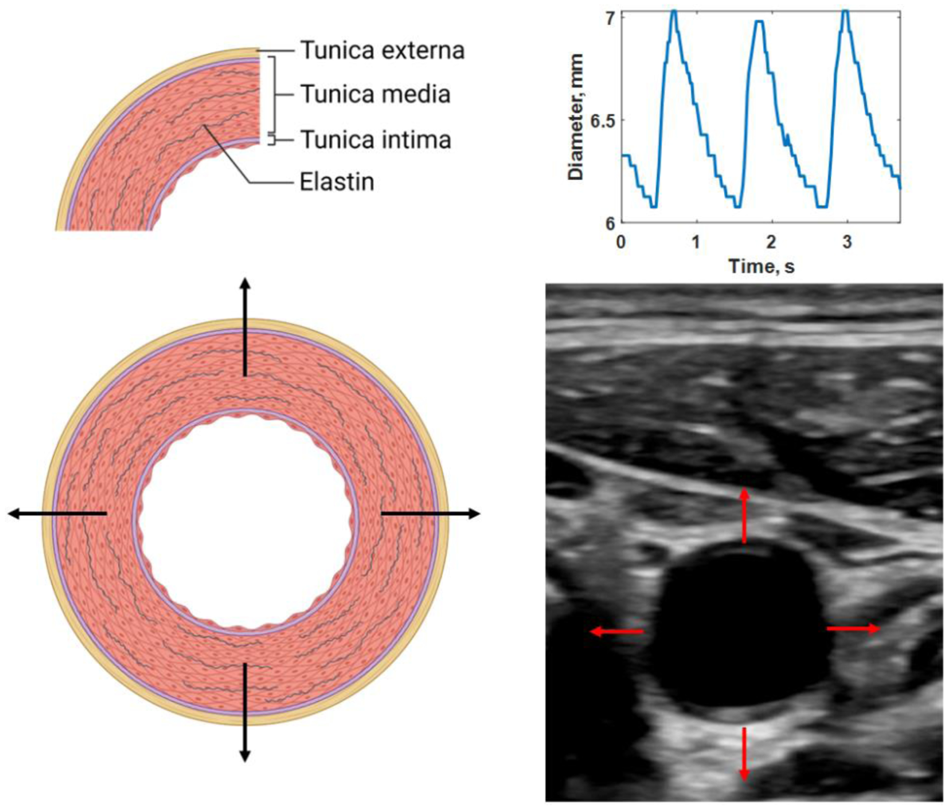
Distension of elastic artery with three layers: tunica externa, tunica media, and tunica intima (A). Ultrasound has a sufficient frame rate to measure real-time motion (B) and, thus the diameter change over the cardiac cycle (C). Created with Biorender.com. See Multimedia1 for a video of the pulsating vessel.