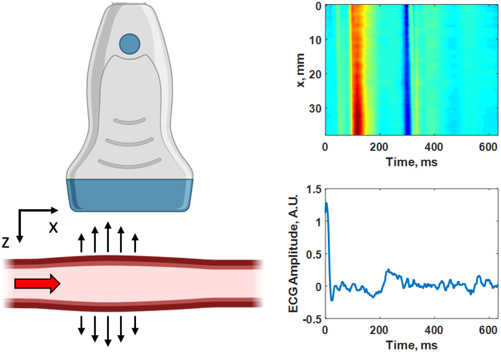
Figure 3.

Pulse wave imaging (PWI) as the pulse travels from left to right (+x direction) passing through the imaging plane (x-z plane) captured by the ultrasound transducer. A diagram of the wall velocity as a function of distance (x-direction) along a human carotid artery is shown on the right with the corresponding electrocardiogram (ECG) trace below with the time axes being the same for both plots. In the upper image, red corresponds to motion towards the transducer and blue corresponds to motion away from the transducer. The PWV is determined by tracking the time-of-arrival of the wave peak in red or other feature at each position along the length of the artery (x-direction), and in this case the PWV = 5.01 m/s. Created with Biorender.com.
