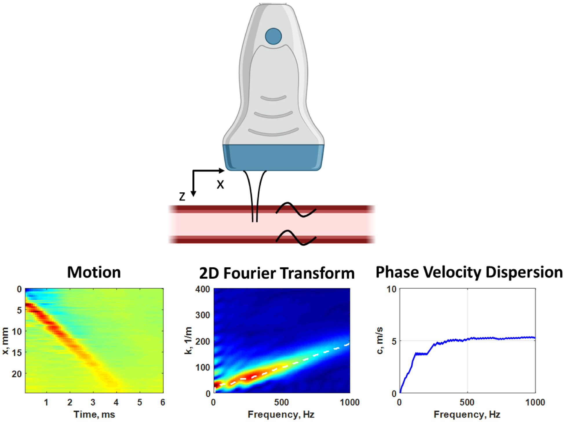
Figure 4.

Measurement with ARF excitation of propagating waves in the x-direction in the human carotid artery, while imaging in the x-z plane with the ultrasound transducer (top). The ARF “tap” causes the wave motion from the top wall of the artery (left). We apply a two-dimensional Fourier transform and examine the magnitude distribution (center). The peaks in the magnitude distribution are identified for each frequency to estimate the phase velocity dispersion curve (right). Created with Biorender.com.
