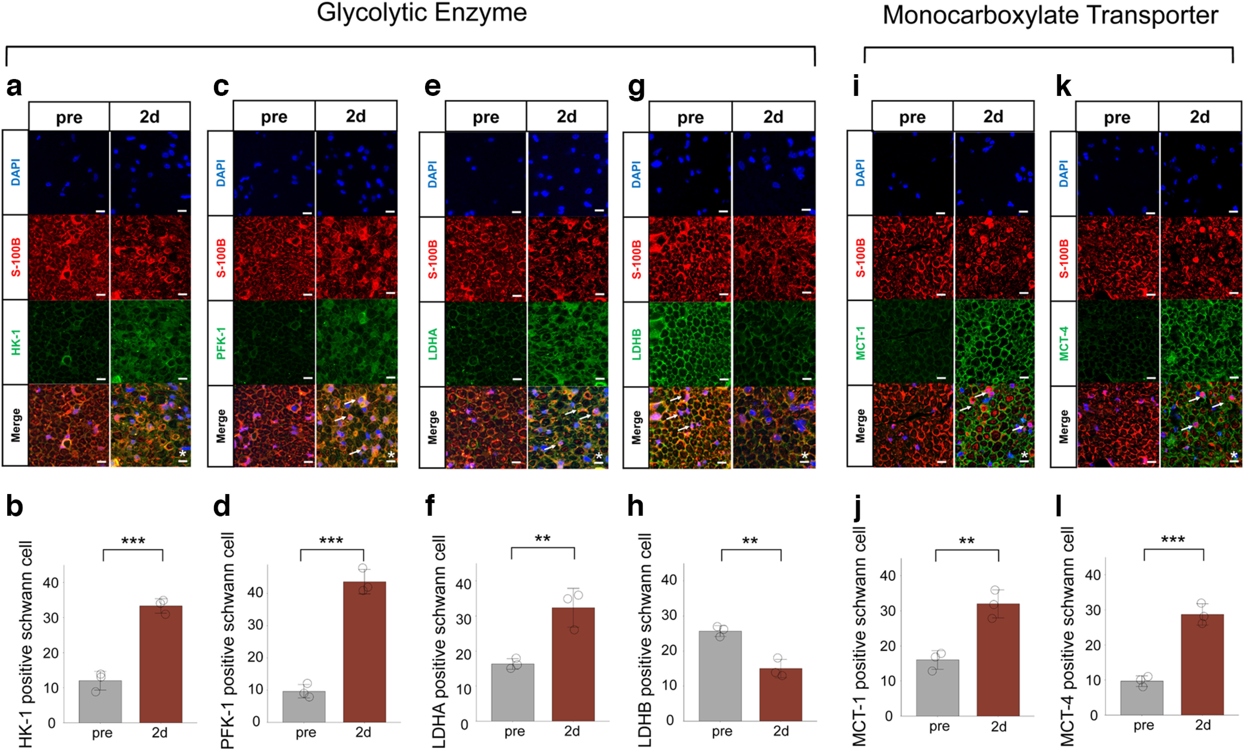
Figure 5.

Activation of the glycolytic system and MCTs in Schwann cell after axotomy. a, c, e, g, Representative cross-section images of immunohistochemistry for glycolytic enzyme (a: HK-1, c: PFK-1, e: LDHA, g: LDHB) before and 2 d after axotomy. b, d, f, h, The number of HK-1 (b), PFK-1 (d), LDHA (f), and LDHB (h) positive Schwann cells (n = 3 mice per group). HK-1, PFK1, and LDHA positive Schwann cells were significantly increased after axotomy, whereas LDHB positive Schwann cells were significantly decreased. i, k, Representative images of immunohistochemistry for MCT (e: MCT-1, g: MCT-4) before and after axotomy. j, l, The number of MCT-1 (j) or MCT-4 (l) positive Schwann cells (n = 3 mice per group). MCT-1 and MCT-4 positive Schwann cells were significantly increased after axotomy. All histologic evaluations were performed 3 mm distal to the sectional end and corresponding uninjured nerve. Error bars indicate SD. Arrows, HK-1/PFK-1/LDHA/LDHB/MCT-1/MCT-4 positive Schwann cells; scale bar, 10 μm. *p < 0.05, **p < 0.01, two-tailed t test. HK, hexokinase; LDHA, lactate dehydrogenase A subunit lactate; LDHB, lactate dehydrogenase B subunit lactate; dehydrogenase; MCT-1, monocarboxylate transporters 1; MCT-4, monocarboxylate transporters 4; PFK, phosphofructokinase.
