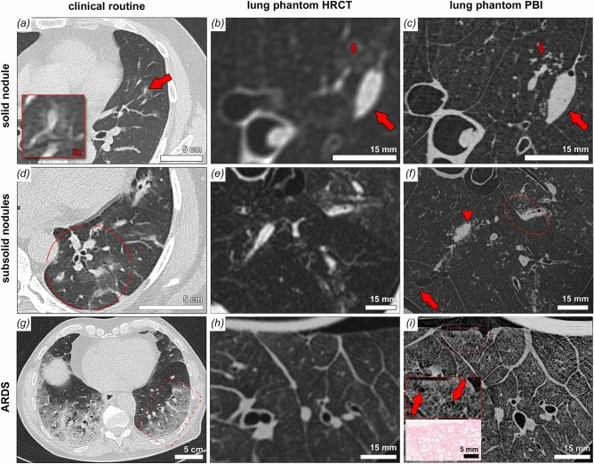
Figure 5.
Correlative cHRCT and PBI SRCT with comparative clinical cases. a, d and g show comparative clinical examples of pulmonary damage patterns from cases of viral pneumonia. Patterns of solid nodules (a), subsolid nodules (b) and acute interstitial pneumonitis/acute respiratory distress syndrome (g) are shown. Artificial lesions mimicking those patterns were imaged by clinical HRCT (b, e and h) and the same lung regions were imaged by PBI-based local tomography (c, f and i). For solid nodules PBI-SRCT proofs superior characterization of the lesions borders and substructures (arrow), note the clear depiction of tiny gas enclosures and the orientation of the nodule alongside an interlobular septum. Also a faint ground glass area (§ in b) is unveiled as micro tree-in-bud (§ in c) by PBI-based SRCT. Grouped subsolid nodules can be depicted in detailed context to the surrounding lung parenchyma. In the circle in (f), an extension of the solid component into the interlobular septum can be noted and tiny substructures of the solid component can be depicted (air enclosures indicated by arrowhead in f). Perifocal micro tree-in-bud patterns can be attributed to the ground glass components and a parenchymal laceration can be noted (arrow in f). In cases of acute alveolar damage (g–i) substructures of the secondary pulmonary lobule become accessible by PBI-based SRCT, note the heterogeneous distension of individual alveolar air spaces (box in h, arrows) with correlative H&E stained histology of the same specimen.

