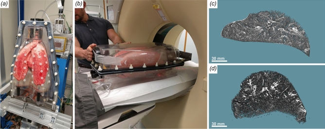
Figure 7.
Comparison of synchrotron CT and clinical CT scanning of the human chest phantom. (a) Phantom mounted in up-right position at the synchrotron setup. (b) Phantom placed in horizontal orientation in the clinical CT scanner. (c) Sample lung region extracted from the synchrotron data. (d) The same lung region (from the same lung) extracted from the clinical CT. Clearly, the shape of that region differs dramatically between the two setups.

