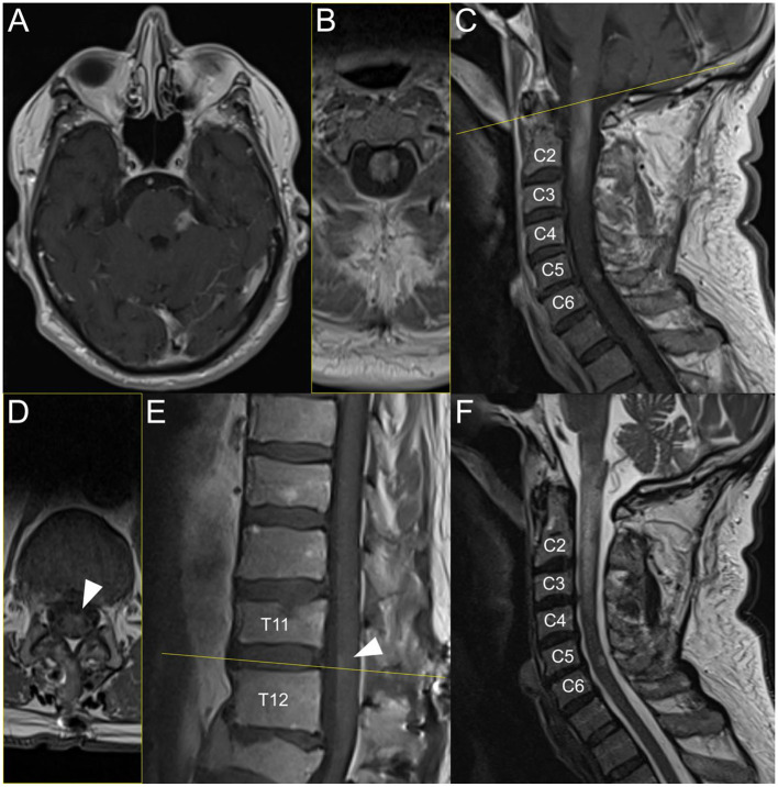
Figure 1.
Brain and spinal MRI during post-SARS-CoV-2 infection-induced ADEM. Acute inflammatory contrast-enhancing T1 MRI lesions on the left middle cerebellar peduncle (A), longitudinal extensive transverse myelitis from the lower part of the medulla oblongata to C3 (C); [panel (B) shows axial plane, i.e., yellow line on panel (C)] with edema lesion on T2 (F) reaching C4, on level C5/C6 (C, F), and on the ventral conus (E); [panel (D) shows axial plane, i.e., yellow line on panel (E)].

