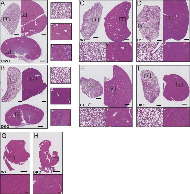
Figure S1.
Organ pathology after systemic infection with S. aureus. (A and B) Representative H&E-stained sections of lungs (1), liver (2), and kidneys (3) from uninfected WT (UIWT) mice (A) and D1−/−D1L3−/− (DKO) mice (B) 18 h after infection with S. aureus. (C–H) Representative H&E-stained sections of lungs (1) and liver (2) from WT (C), D1−/− (D), D1L3−/− (E), and D1−/−D1L3−/− (DKO; F) and heart from WT (G) and D1−/−D1L3−/− (H) mice 72 h after infection with S. aureus. Shown are low-magnification images of organs (scale bar, 1 mm) and high-magnification images of areas highlighted in black rectangles (scale bar, 100 μm).

