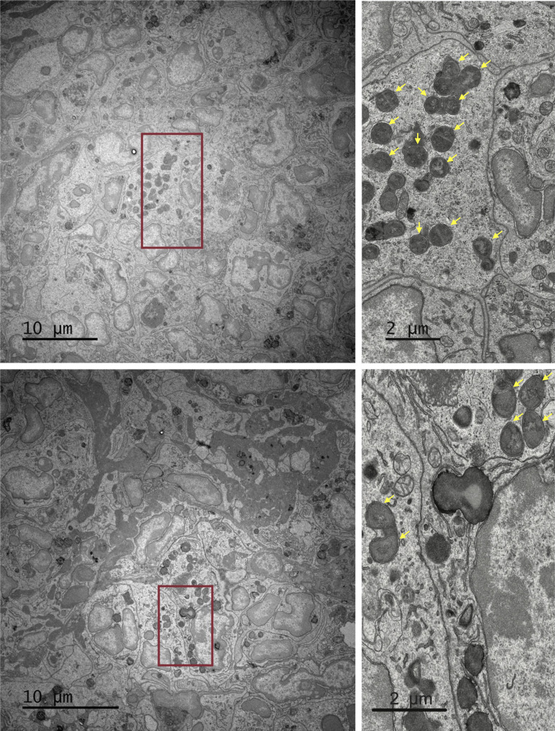
Figure S4.
Detection of S. aureus in the kidney by transmission EM. Shown are representative transmission EM images of kidneys from D1−/−D1L3−/− mice 72 h after infection with 1 × 10⁷ CFU of S. aureus (scale bars, 10 μm). Areas highlighted in red rectangles are shown at higher magnification on the right (scale bars, 2 μm). The S. aureus cells, represented by dense round structures circumscribed by capsules and with an occasional internal septum, are highlighted by arrows.

