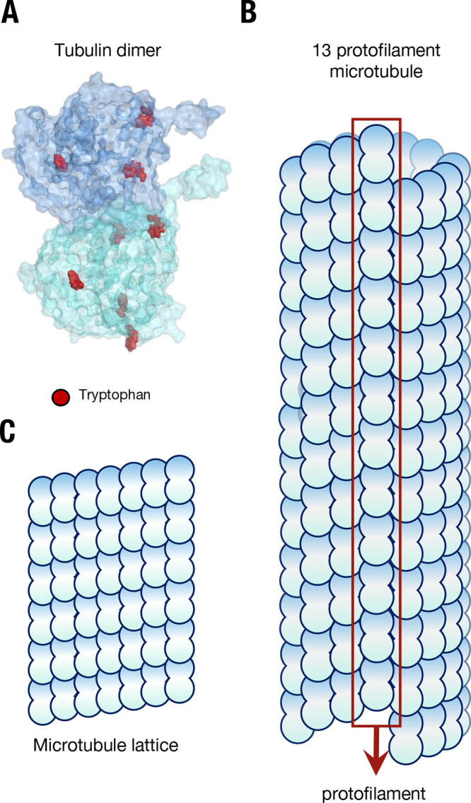
Figure 1.

The structure of microtubules from a lattice of tubulin. (A) The tubulin dimer with tryptophan residues marked in red; the C-termini “tails” can be seen protruding from each monomer. (B) The structure of a microtubule, showing constituent arrangement of tubulin dimers, and the presence of a “seam”. (C) The repeating “lattice” of tubulin dimers in a microtubule.
