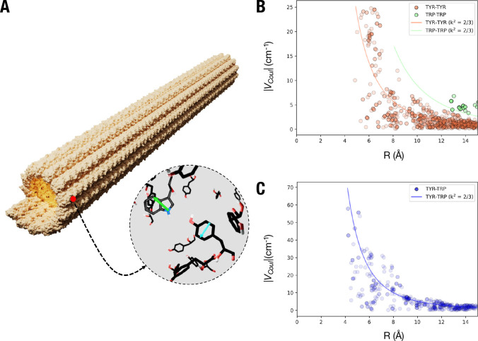
Figure 6.
Theoretical estimation of interactions among tryptophan and tyrosine residues. (A) Crystal structure of a microtubule composed of 31 tubulin dimers stacked vertically showing the dipole moment orientations of representative tryptophan (green; TRP) and tyrosine (cyan; TYR) residues. (B) Distribution of the coupling constant VCoul between TYR-TYR, TRP-TRP and (C), TYR-TRP residues in 31-dimer long microtubule crystal structure. A projection of VCoul with orientation factor (κ2) of 2/3 is represented as solid lines.

