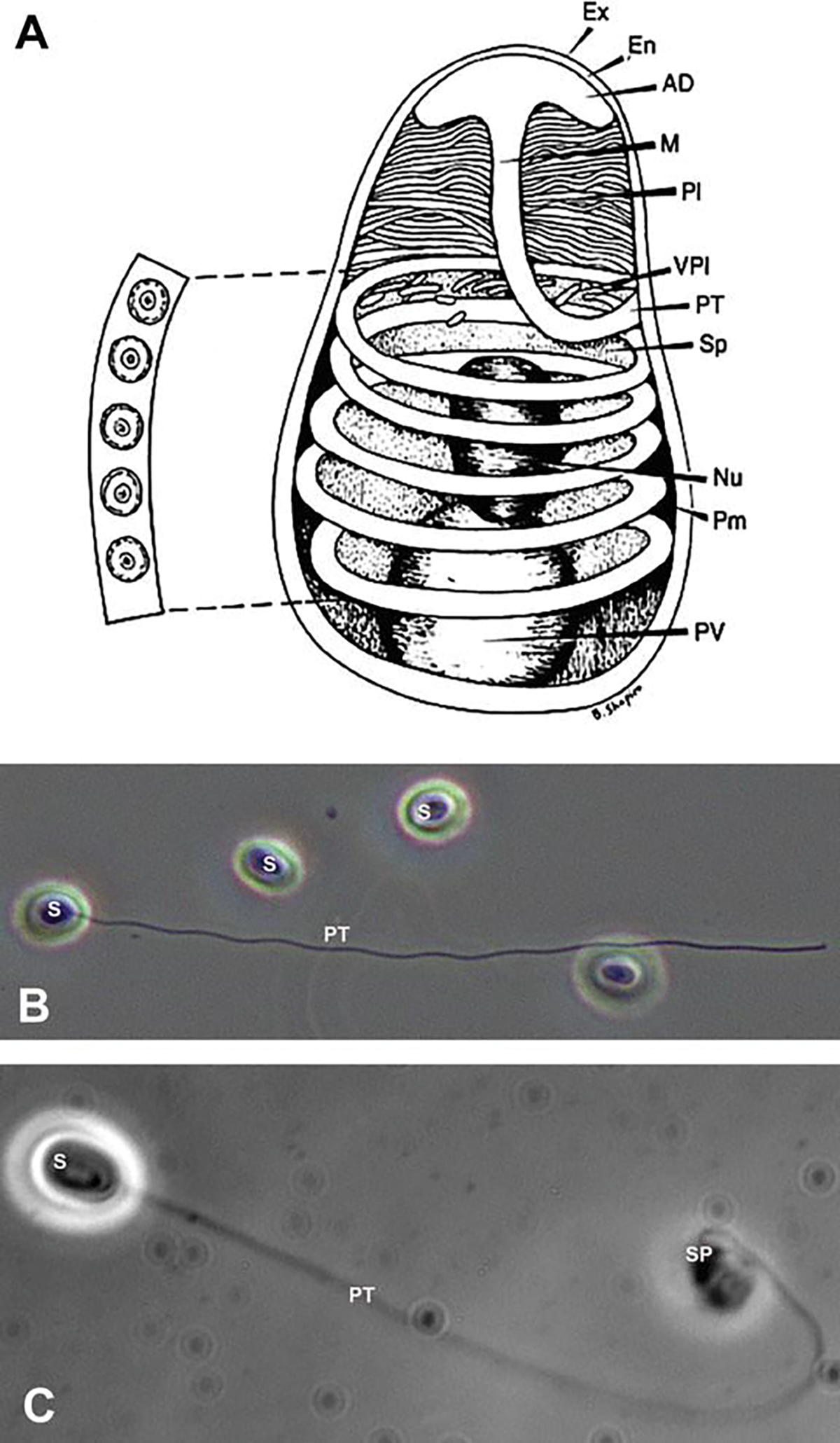
Fig. 8.1.

Microsporidian spore structure and light microscope images. (a) Diagram of a microsporidian spore. Microsporidian spores vary in size from 1 to 12 μm. The spore coat is thinner at the anterior end of the spore and consists of an electron lucent endospore (En), electron-dense exospore (Ex), and the plasma membrane (Pm). The sporoplasm (Sp) contains ribosomes, the posterior vacuole (PV), and a single nucleus (Nu). The anchoring disc (AD) at the anterior end of the spore is the site of attachment of the polar tube. It should be noted that the polar tube is often called the polar filament when it is within the spore prior to germination. The anterior or straight region of the polar tube that connects to the anchoring disc is called the manubroid (M), and the posterior region of the tube coils around the sporoplasm. The number of coils and their arrangement (i.e., single row or multiple rows) is used in microsporidian taxonomic classification. The lamellar polaroplast (Pl) and vesicular polaroplast (VPl) surround the manubroid region of the polar tube. The insert depicts the polar tube coils in this figure in a cross section illustrating that the polar tube within the spore (i.e., polar filament) has several layers of different electron density by electron microscopy. Reprinted with permission from Keohane EM, Weiss LM. 1999. The structure, function, and composition of the microsporidian polar tube. pp. 196–224. In Wittner M, Weiss LM (ed), The Microsporidia and Microsporidiosis. ASM Press, Washington, DC (Keohane and Weiss 1999). (b) Differential interference contrast (DIC) microscopy image of Anncaliia algerae spores. One of the spores (S) has become activated, and the polar tube (PT) is in the process of extrusion. (c) Phase contrast microscopic image of an Anncaliia algerae spore (S) with the extruded polar tube (PT) and the sporoplasm (SPM) still attached to the distal end of the PT
