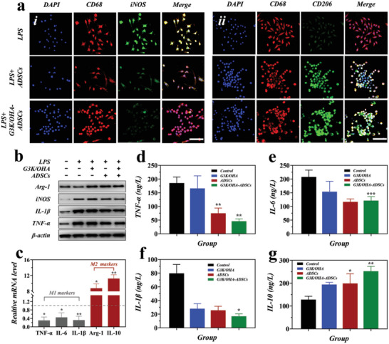
Figure 4.

In vitro M1 to M2 polarization of BMDMs macrophages by G3K/OHA‐ADSCs hydrogels. a) Immunofluorescence staining of CD68 (the pan‐macrophage marker, red), iNOS (M1 marker, green), and CD206 (M2 marker, green), and nuclei (blue) on macrophages without or with the treatment of G3K/OHA‐ADSCs hydrogels. Scale bar = 50 µm. b) Protein expression of M1 (iNOS, IL‐1β, and TNF‐α) and M2 (Arg‐1) macrophage markers in BMDMs under various conditions, as evaluated by Western blot analysis. c) The mRNA levels of M1 macrophage markers (IL‐1β, IL‐6, and TNF‐α), and M2 macrophage markers (Arg‐1 and IL‐10) in activated macrophages without and with the treatment of G3K/OHA‐ADSCs hydrogels, as evaluated by qRT‐PCR analysis. d–g) Supernatant cytokines were detected in BDMDs treated by G3K/OHA, ADSCs, and G3K/OHA‐ADSCs. n = 3. Data were presented as mean ± SD. Statistical significance was calculated by one‐way ANOVA followed by post hoc tests, *0.01 < P < 0.05, **0.001 < P < 0.01, ***P < 0.001.
