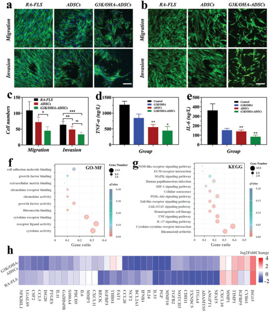
Figure 5.

G3K/OHA‐ADSCs inhibited RAFLS migration, invasion, and inflammatory responses in vitro. a) Actin/diamidino phenylindole (DAPI) and b) Calcein acetoxymethyl (AM) staining of RAFLS in migration and invasion assays. The scale bar is 200 µm. c) Quantification of RAFLS cells migrated and invaded through Trans well. d,e) Supernatant proinflammatory cytokines were detected in RAFLS treated by G3K/OHA, ADSCs, and G3K/OHA‐ADSCs. f) GO and g) KEGG pathway analyses of the target genes of the top ten significantly expressed miRNAs in the ADSCs group compared with the NC group. The GO terms and KEGG pathway terms enriched in the predicted target genes of the miRNAs were analyzed using Database for Annotation Visualization and Integrated Discovery (DAVID) Bioinformatics. MF, molecular functions. h) The expression of marker genes in ADSCs and negative control (NC) groups. n = 3. Data were presented as mean ± SD. Statistical significance was calculated by one‐way ANOVA followed by post hoc tests, *0.01 < P < 0.05, **0.001 < P < 0.01, ***P < 0.001.
