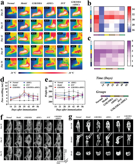
Figure 6.

In vivo assessments of pathological features of CIA rat models after intra‐articular injection of various materials. a) Thermographic images of left hind paws and corresponding quantification of b) paw temperatures, c) clinical scores, d) paw swelling, and e) body weight at various time points after treatment. f) Representative 3D reconstructed micro‐CT images of the knee joints and corresponding 2D images in the COR, SAG, and AX planes. n = 5, biologically independent samples. Data were presented as mean ± SD.
