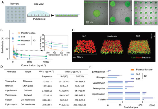
Figure 2.

Mechanical rigidities promoted antibiotical resistance of bacterial microcolonies in the developed in vitro 3D AST model. A) Schematic diagram of a PDMS microwell array for in vitro 3D AST. The three sub‐images on the right showed representative ones of bacterial microcolonies cultured in the PDMS microwell arrays whose depths were 300 µm while the corresponding diameters were 200, 800, and 1000 µm, respectively. Note that the bacterial microcolonies were beforehand labeled as green in experiments. Scale bar: 400 µm. B) Quantitative dependence of survival rates of planktonic E. coli, the corresponding microcolonies grown in soft, moderate, and stiff MA hydrogel samples on concentrations of broth microdilution erythromycin (0‐10 000 µg mL−1). C) Results of live/dead staining for E. coli microcolonies in soft, moderate, and stiff MA hydrogels, respectively, after treatments with 5000 µg mL−1 erythromycin. The live and dead bacterial cells were labeled green and red, respectively. Scale bar: 50 µm. D) MICs and MBECs of planktonic E. coli and the corresponding microcolonies cultured in soft and stiff MA hydrogels after treatments with various antibiotics with distinct targets. Data were presented as mean±SD from at least three individual experiments. E) Ratios of MBECs of bacterial biofilms grown in soft and stiff MA hydrogels to MIC of the corresponding planktonic bacteria after they were treated with various antibiotics.
