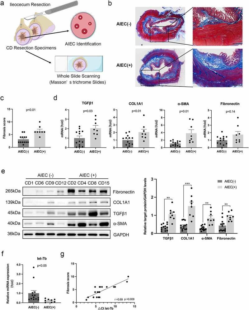
Figure 1.

AIEC colonization is associated with the enhanced intestinal fibrosis in CD patients. (a) Illustration of the disposition of surgical resection specimens from CD patients. The slides of terminal ileum specimens are subjected to whole slide scanning to evaluate fibrosis after Masson’s trichrome staining, and mucosal samples are collected from the same sites of the specimens for AIEC identification. (b) Collagen fiber deposition in the submucosa and muscularis propria areas in AIEC(+) and AIEC(-) CD patients presented by Masson’s trichrome staining. Scale bar, 1 mm. (c) Fibrosis scores for quantification of degrees of intestinal fibrosis in AIEC(-) and AIEC(+) CD patients. (d&e) The mRNA and protein levels of TGFβ1, COL1A1, α-SMA and fibronectin in the terminal ileal tissues of AIEC(+) and AIEC(-) CD patients analyzed by qPCR and western blot, respectively. (f) The miRNA levels of let-7b analyzed by qPCR. U6 was used as an internal control. (g) Correlation between the levels of let-7b and fibrosis score in the terminal ileum. Spearman’s test was used for correlation analysis. AIEC(-): AIEC-negative colonization (n = 13); AIEC(+): AIEC-positive colonization (n = 8); data are expressed as the means ± SEM.
