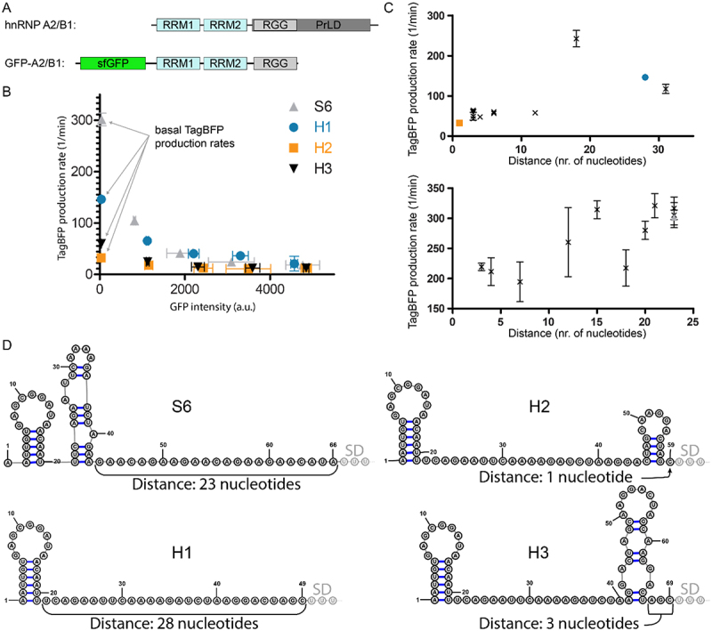
Figure 4.

A) Schematic representation of hnRNP A2/B1 and the GFP-A2/B1 protein construct. B) Translational repression assay results for RNA reporters H1-H3 in the presence of GFP-A2/B1 in comparison with RNA reporter S6 in the presence of GFP-SRSF1 C) Correlation between TagBFP production rate and the distance between a secondary structure and the SD sequence for GFP-A2/B1 (top) and GFP-SRSF1 (bottom). D) Predicted secondary structures of the inserted RBP binding sequences for 5’-UTRs of the reporters S6 and H1-H3 using RNAfold. The first three nucleotides of the SD sequence are indicated as well as the distance between the SD and the closest predicted secondary structure.
