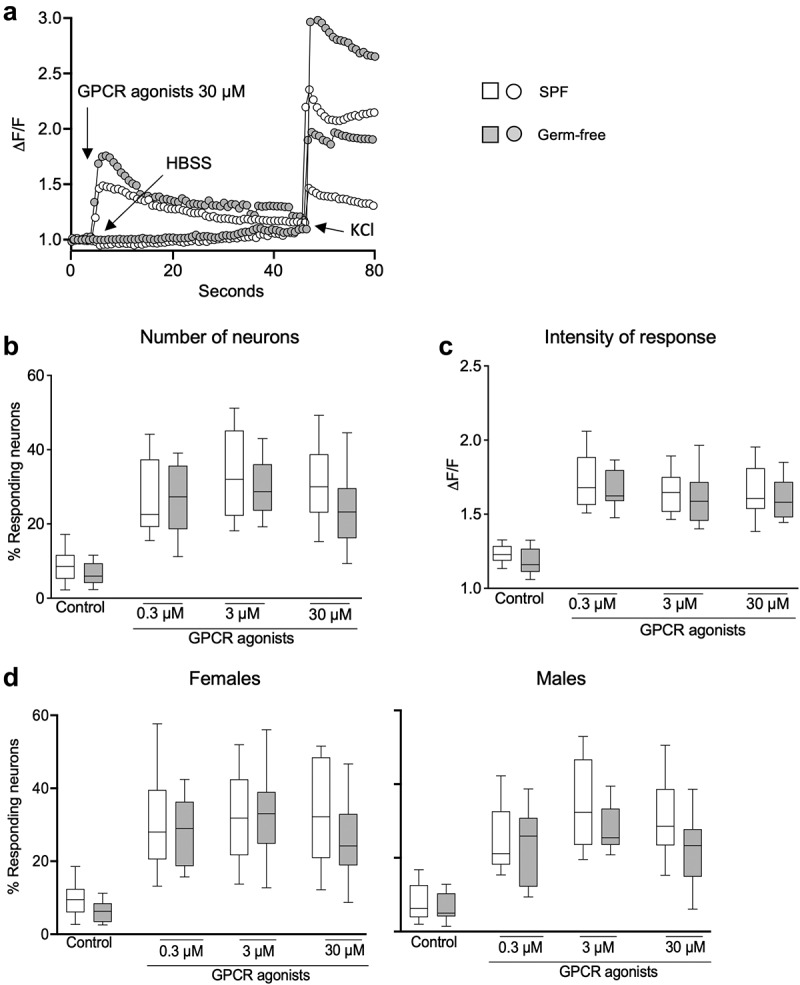
Figure 5.

DRG neuronal activation is similar in SPF and GF mice after GPCR activation. (a) Representative fluorescent traces of calcium flux in DRG neurons from SPF and GF mice in response to vehicle (HBSS) or GPCR agonists (30 μM). (b) Percentage of responding DRG neurons obtained from SPF (white box) and GF (gray box) mice, after treatment with GPCR agonists (0.3 μM, 3 μM, 30 μM). (c) Intensity of the neuronal response (ΔF/F) in DRG neurons obtained from SPF and GF mice, after treatment with GPCR agonists. (d) Percentage of responding neurons obtained from SPF females and males and from GF female and male mice, after treatment with capsaicin. White box: SPF, gray box: GF. Data are represented as box plots (10–90%ile) with n = 8 independent experiments of 1–2 wells per condition for SPF females; n = 7 independent experiments of 1–2 wells per condition for SPF males; n = 5 independent experiments for both GF females and males mice. In each well, 20–130 neurons were cultured. Statistical analysis was performed using 2-way ANOVA followed by šidak’s multiple comparisons test.
