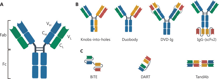
Figure 1.
The immunoglobulin G (IgG) structure and schematic diagram of several representative bsAbs. The IgG is roughly “Y-shaped”. The two heavy chains are shown in blue and the two light chains are shown in green (A). IgG-like bsAbs (B). Non-IgG-like bsAbs (C). BiTE, bispecific T-cell engager; DART, dual-affinity retargeting molecule; DVD-Ig, dual-variable-domain immunoglobulin; scFv, single-chain variable fragment; TandAb, tandem diabody.

