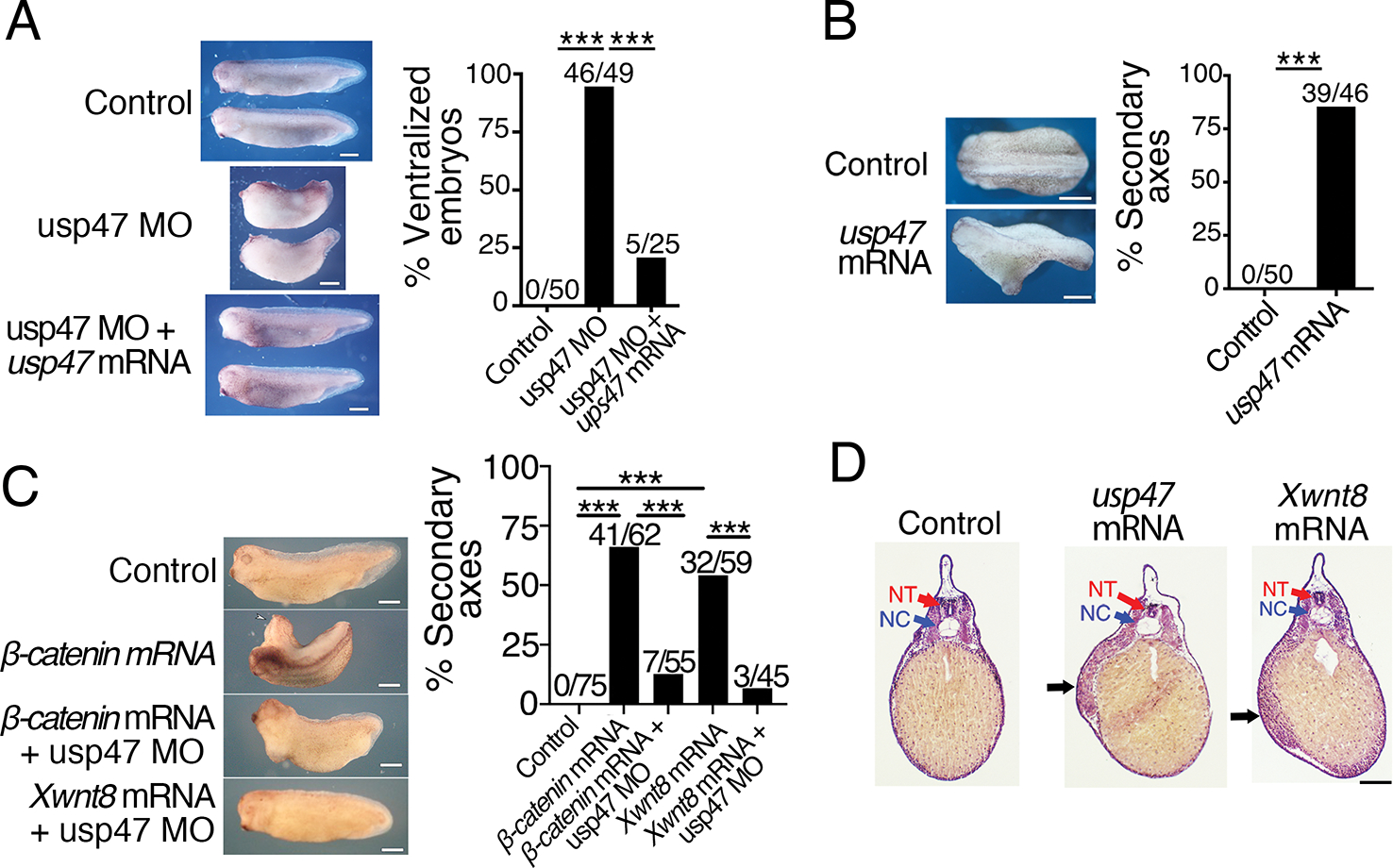
Fig. 2. Loss and gain of Usp47 perturbs axis formation in Xenopus embryos.

(A) Xenopus embryos at the 4-cell stage were injected dorsally with control morpholino, usp47 Morpholino (MO), or usp47 MO plus usp47 mRNA, and the dorsal-anterior index (DAI) was determined as previously described (53). The percentage of ventralized embryos (DAI ≤ 2) was quantified, and absolute numbers are shown above the bars in the graph. Representative embryos are shown. n=25–50 embryos per treatment group in each of 3 independent experiments. ***p < 0.0005. Significance was assessed using Chi-square test with Bonferroni correction. Scale bar, 1 mm. (B) 4-cell embryos were injected ventrally with control or usp47 mRNA. The percentage of embryos with secondary axis formation was quantified, and absolute numbers are shown above bars in the graph. Representative embryos are shown. n= 45–50 embryos per treatment group in each of 3 independent experiments. ***p < 0.005. Significance was assessed using Chi-square test. Scale bar, 1 mm. (C) Xenopus embryos were injected with control MO, β-catenin mRNA, XWnt8 mRNA, β-catenin mRNA plus usp47 MO, or XWnt8 mRNA plus usp47 MO. The percentage of embryos with secondary axis formation was quantified, and the absolute numbers are shown above bars in the graph. Representative embryos are shown. n= 45–75 embryos per treatment group in each of 3 independent experiments. **p < 0.0003. Significance was assessed using Chi-square test with Bonferroni correction. Scale bar, 1 mm. (D) Embryos injected with control, usp47, or Xwnt8 mRNA were fixed, sectioned, and stained with hematoxylin and eosin. Black arrows point to ectopic mesoderm. NT, neural tube; NC, notochord. Scale bar, 200 μm. Images are representative of n > 5 embryos per treatment group.
