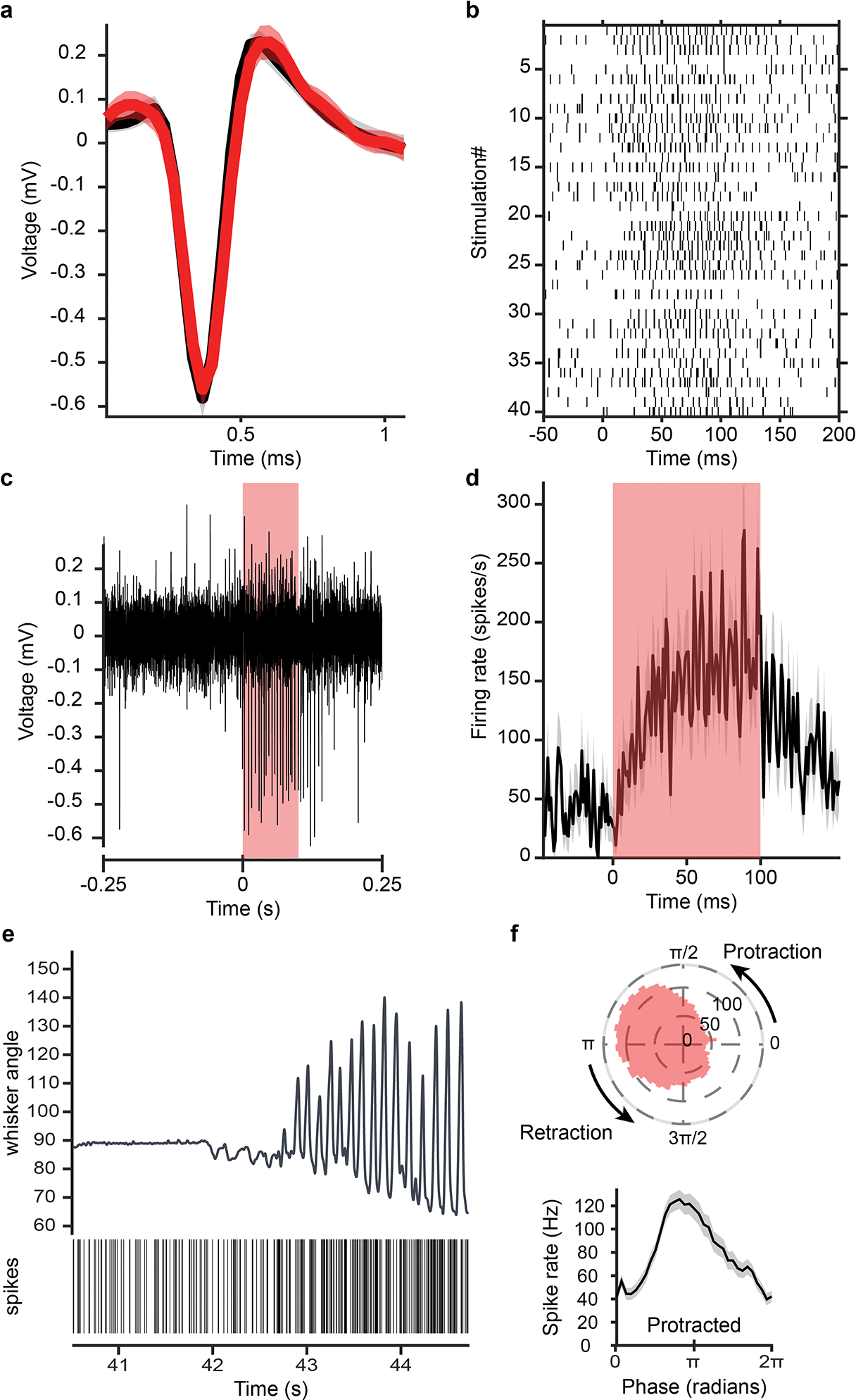
Extended Data Fig. 3. Antidromic opto-tagging of a ChRmine-expressing vIRtPV neuron via light stimulation through the ear.

a, Overlapped average waveform, before (black) and during (red) stimulation periods. b, Raster plot of spike times aligned to stimulation onset for 40 light pulses. c, Example single channel recording trace showing antidromic spikes from the opto-tagged unit during a light pulse. d, Firing rate of that unit averaged over 40 light stimulations epochs. e, Unit activity during transition from resting to whisking state. vibrissa angle traces for the ipsilateral C2 vibrissa. Bottom: Spike raster plot for this opto-tagged vIRtPV-ChRmine neuron. f, Phase tuning. Average spike rate across whisking phases for this opto-tagged vIRtPV-ChRmine neuron, in polar (top) and cartesian coordinates (bottom).
