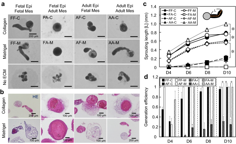
Figure 2.
Characterization of human hair follicloids. (a) Self-organization of the epithelial and mesenchymal cells in hair follicloids. Hair follicloids were prepared by combining fetal and adult epithelial and mesenchymal cells, supplemented with or without collagen and Matrigel (Table 1). Stereomicroscope images were observed on day ten of culture. (b) Cross-sectional view of hair follicloids. On day ten of culture, hair follicloids were sectioned and stained with hematoxylin and eosin. (c) Length of the sprouting structures generated from hair follicloids prepared under different conditions (Table 2). The length was measured from the stereomicroscope images taken on days four, six, eight, and ten of culture. (d) Generation efficiency of sprouting structures after day ten of culture. The average ratio was analyzed from the results of three independent experiments.

