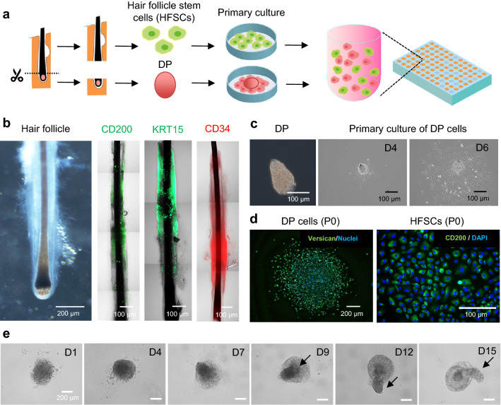
Figure 5.
Preparation of hair follicloids using patient hair follicle-derived cells. (a) Preparation of hair follicle stem cells (HFSCs) and dermal papilla (DP) cells from hair follicles of patients with androgenic alopecia. Dissociated HFSCs and DP tissues were seeded on the dishes for primary culture. Proliferated HFSCs and DP cells were suspended in culture media containing Matrigel and cultured in a 96-well plate. (b) Native human hair follicles. Microscope and immunostaining images were taken before cell dissociation. (c) DP cell culture. Isolated DPs adhered to and proliferated on the culture dishes. (d) Expression of stem cell markers in HFSC and DP cells. Primary cultures containing HFSCs and DP cells were visualized through the immunohistochemical staining of versican and CD200. (e) Time-course of sprouting of hair follicloids.

