
Figure 4.
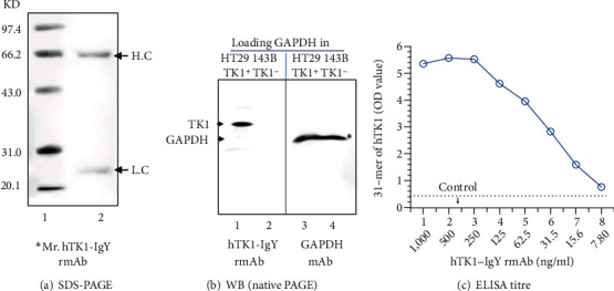
Specificity and sensitivity of hTK1-IgY-rmAb#5: (a) SDS-PAGE detection of the purified antibody (0.5 μg). The IgY molecule heavy chain (H) was ≈66 kDa, and the light chain (L) was ≈25 kDa. (b) Western blot detection of antibody specificity; TK1-negative cell lines (143BTK1−) and TK1-positive cell lines (HT29) were used, and GAPDH was used as a loading control. (c) ELISA titre detected the #5 antibody. ∗Mr.: molecular weight marker.