
Figure 2.
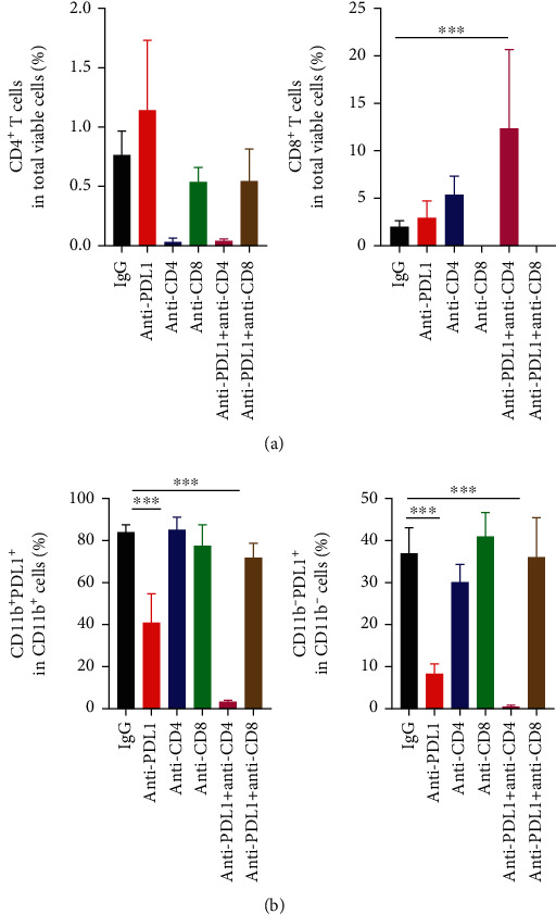
Anti-PD-L1 therapy decreased the proportions of intratumoral PD-L1+ immune cells in a CD8+ T cell dependent manner, while CD4+ T cells antagonized those effects. CT26 tumor-bearing mice were prepared and treated as described in Figure 1. On day 12 post anti-PD-L1 treatments, tumor tissues were harvested and analyzed by flow cytometry. (a) The proportions of intratumoral CD4+ and CD8+ T cells were analyzed by flow cytometry. (b) The percentages of intratumoral PD-L1+CD11b− cells in lymphoid cells and PD-L1+CD11b+ cells in myeloid cells were assessed by flow cytometry. Significant differences were determined by one-way ANOVA. Data were from one experiment representative of two independent experiments with similar results (n = 7 − 8 mice per group). Data were shown as means ± SD. ∗P < 0.05, ∗∗∗P < 0.001.