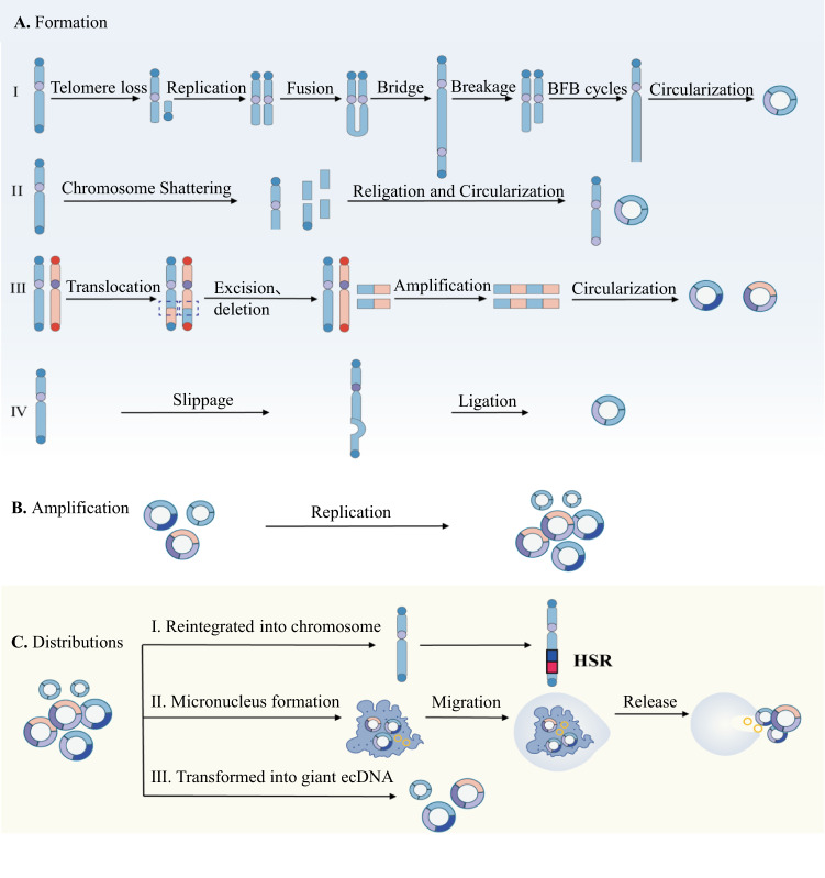
Fig. 1. Biogenesis, amplification and distribution of ecDNA.
A Formation of ecDNA. (I) Breakage-fusion-bridge (BFB) cycles. Loss of telomere because of genome instability, and the end of the missing telomere fuse with each other to form a chromosomal structure with two centromeres and a dicentric anaphase bridge. The fusion bridge is broken in the late stage of mitosis, keep the genes amplified and circularizing into ecDNA. (II) Chromothripsis model. When chromosomes are catastrophically broken, the DNA double-strand break into some DNA segments, which are randomly linked and cycled to form ecDNA during subsequent DNA repair. (III) Translocation-excision-deletion-amplification (TEDA) model. Segments between chromosomes translocation, DNA fragments between translocation breakpoints are prone to amplification, retention or deletion, and the deleted part is cyclized outside the chromosomes to form ecDNA. (IV) Episome model. Through the way of DNA slippage and R-loop, chromosomes form episomes during genetic recombination, ecDNA generated by cleavage and ligation. B Amplification of ecDNA replicates by rolling circle amplification. C ecDNA distributions. ecDNA can be subject to further clonal evolution, reintegrated into chromosomes, combined with other ecDNAs or eliminated by being trapped inside micronucleus.

