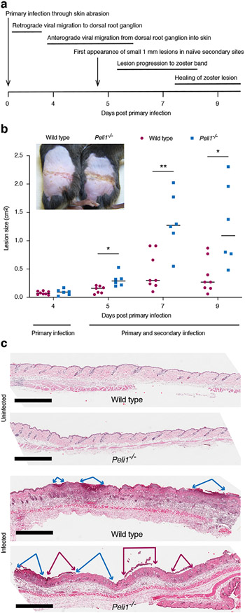
Figure 2: HSV-1 causes larger lesions in Peli1−/− mice.
Wild type and Pellino-1 KO mice were infected with HSV-1. (a), Timing and progression of HSV infection in the mouse flank model is illustrated. (b), Lesions on the flanks were photographed up to day 9 and sized using ImageJ. Representative data and images from one of at least 3 independent experiments (n = 4-5 per group) is shown. Each dot represents a single mouse. *, p < 0.05; **, p < 0.01 (t test), (c), Skin area with early zosteriform sites above and below the primary infection site were collected 5 days post-primary infection and histology evaluated alongside uninfected skin by H&E staining. Scale bar = 1 mm. Blue and magenta arrows mark regions with absent and detaching epidermis, respectively.

