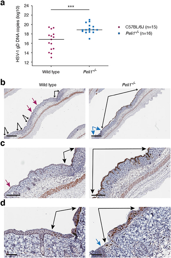
Figure 3: Pellino1 limits viral replication in the epidermal keratinocytes.
Five days post-HSV-1 infection, skin was collected from above and below the primary infection site and examined by QPCR for HSV-1 DNA (a) or immunohistochemistry for HSV-1 protein (b). (a), Each symbol represents a single mouse. Data is pooled from 3 independent experiments. ***, p < 0.001 (t test), (b-d), Black arrows flank continuous epidermal regions with HSV-1 positive staining. Blue arrows flank lesion site with absent epidermis. Magenta arrows identify small HSV-1 positive foci. Larger magnifications of images in (b) are shown in (c) and (d). Scale bars represent 700 μm (b), 300 μm (c), and 70 μm (d), respectively. Images are representative from 2 independent experiments each involving 4-5 mice per group.

