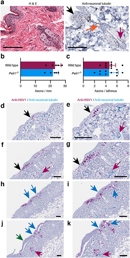
Figure 5: HSV-1 disseminates from the surface epidermis through the infundibulum to the sebaceous gland.
Uninfected and HSV-1 infected skin was examined by H&E and single (a-c) or dual (d-k) immunohistochemistry for HSV-1 (magenta, d-k) and/or neuronal tubulin (brown, a; turquois, d-k). (c) Number of axons crossing the dermal/epidermal boundary (b) and wrapped around the isthmus. Images in e, g, i and k are larger magnifications of images in d, f, h and j, respectively. Black arrows identify free nerves in the epidermis. Orange and magenta arrows point to mechanosensory nerves wrapped around the infundibulum and isthmus, respectively. Blue arrows indicate positions of infected infundibulum. Green arrow points to infected epidermis where most infected keratinocytes have already died. Scale bars represent 50 μm.

