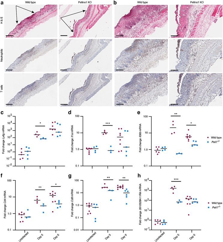
Figure 6: Recruitment of immune cells is delayed in Pellino1 KO mice.
(a) Early zosteriform lesion areas above and below the primary infection site were collected at day 5 post-primary infection from wild type and Pellino1 KO mice. The skin was examined by H&E and immunohistochemical staining for neutrophils and T cells. Representative images focused on skin regions with approximately equally wide involvement of the epidermis are shown flanked by black arrows (a). Larger magnifications of images in (a) are shown in (b). Scale bars represent 500 μm (a) and 200 μm (b), respectively. (c-h) Wild type (red circles) and Pellino1 KO (blue squares) mice were left uninfected or infected with HSV-1, and skin collected at the indicated times. On day 5, 1 mm secondary lesions were collected with 4 mm punch biopsies. On day 6, 8 mm punch biopsies across the entire secondary regions were collected. Expression of Ly6g (c), Il1a (d), Il36b (e), Cd4 (f), Cd8 (g), and 2610528A11Rik (Gpr15l) (h) mRNAs were examined by RT-PCR. Each symbol represents a single mouse. Data for each timepoint is representative of 3 independent experiments. *, p < 0.05; **, p < 0.01; ***, p < 0.001 (t test).

