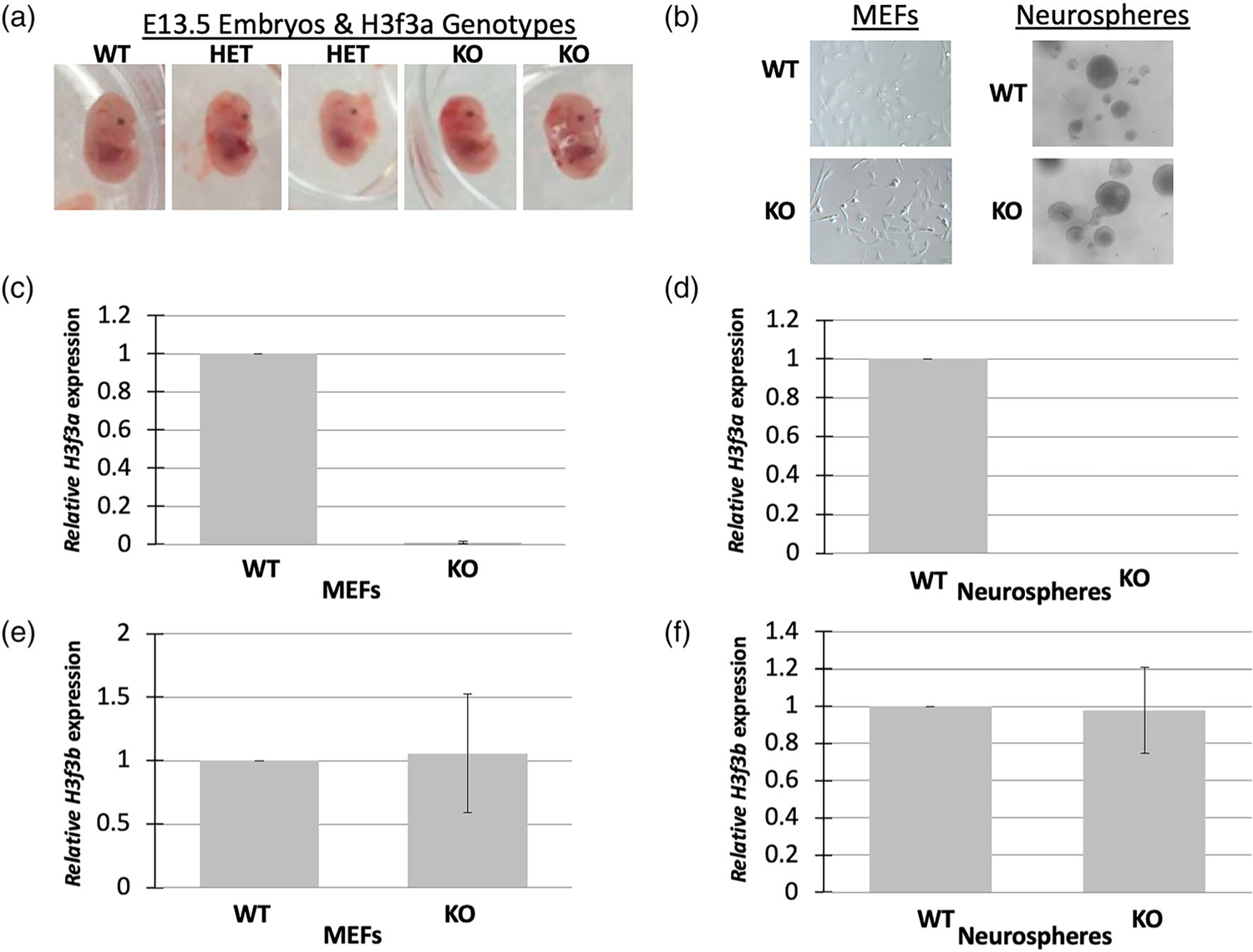
FIGURE 3.

H3f3a targeted MEFs and neurospheres. (a) Heterozygous and null mid-gestational embryos exhibit aberrant phenotypes including small heads. Timed matings were conducted and E13.5 embryos were isolated from heterozygous crosses. Representative embryos are shown. (b) MEFs and neurospheres were isolated from the embryos and cultures established. There were no obvious phenotypes of the cells of different genotypes. (c–d) H3f3a null MEFs and neurospheres exhibited nearly undetectable levels of H3f3a RNA, and (e–f) no significant changes in H3f3b RNA. MEFs and neurospheres were produced from one WT, two heterozygous, and two null embryos for RNA analysis
