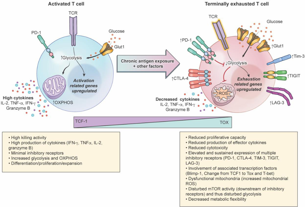
Figure 1.
Characteristics of functional and terminally exhausted T-cells. Chronic antigen exposure and other intrinsic and extrinsic factors drive T-cell exhaustion in cancer. Expression of inhibitory receptors CTLA-4, PD-1, TIM-3, TIGIT, and LAG-3 increases in exhausted T-cells compared to functional T-cells; transition to an exhausted state is also marked by a decrease in TCF-1 expression and increase in TOX expression. Disturbed TCR signaling, Glut1 upregulation, elevation of inhibitory receptors, and transcriptional and epigenetic changes drive metabolic dysfunction, impairing glycolytic abilities and metabolic flexibility in exhausted T-cells. Cytokines, including IL-2, TNF-α, and IFN-γ, and effector protease granzyme B production also decrease in exhausted T-cells compared to functional ones. These alterations, along with reduced proliferative capacity, results in diminished cytotoxicity as T-cells progress to terminal exhaustion.

