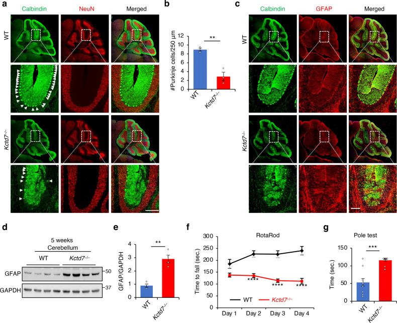
Fig. 7. KCTD7 deficiency leads to behavioral impairment and neurodegeneration.
a–c Confocal microscopy analysis of the cerebellum from 5-week-old WT and Kctd7–/– mice. α-calbindin antibody and α-GFAP antibody were used to label Purkinje cells (a) and astrocytes (c), respectively. Quantification of Purkinje cell numbers is reported in b, n = 5 per genotype. Scale bars, 200 μm. d, e Immunoblot analysis of cerebellar tissue from 5-week-old WT and Kctd7–/– mice using α-GFAP antibody. f Rotarod test measuring the latency to fall for 8-week-old WT (n = 10) and Kctd7–/– (n = 16) mice. g Inverted pole test measuring the time used to climb down from the pole for 8-week-old WT (n = 10) and Kctd7–/– (n = 19) mice. Data represent means ± SEM; ∗P < 0.05, ∗∗P < 0.01, ∗∗∗P < 0.001, ns not significant.

