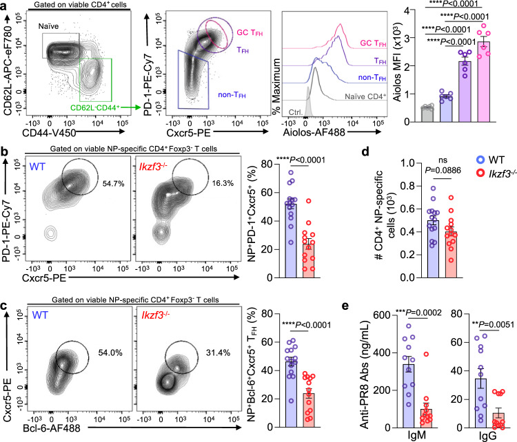
Fig. 1. Aiolos expression is elevated in TFH cell populations, and its loss results in disrupted TFH cell differentiation and antibody production in response to influenza infection.
a Naïve WT C57BL/6 mice were infected intranasally with 30 PFU influenza (A/PR8/34; “PR8”) for 8 days. Single-cell suspensions were generated from the lung-draining lymph nodes (DLN), and analysis of Aiolos protein expression in the indicated populations was performed via flow cytometry. Data are compiled from 2 independent experiments (n = 6 ± s.e.m; ****P < 0.0001; one-way ANOVA with Tukey’s multiple comparison test). b–d Analysis of the percentage of influenza nucleoprotein (NP)-specific PD-1HICxcr5HI (TFH) populations generated in response to influenza infection. Following single-cell suspension, cells were stained with fluorochrome-labeled tetramers to identify NP-specific populations. b, c Analysis of the percentage of influenza nucleoprotein (NP)-specific Bcl-6HICxcr5HI (TFH) populations generated in response to influenza infection (For ‘b’, n = 14 for WT and 13 for Aiolos KO. For ‘c’, n = 14. Data are presented as mean ± s.e.m; data are compiled from four independent experiments; ****P < 0.0001; two-sided, unpaired Student’s t test). d Total NP-specific CD4+ T cells generated in WT versus Aiolos-deficient animals following influenza infection were enumerated. (n = 14 for WT and 13 for Aiolos KO. Data are presented as mean ± s.e.m; data are compiled from 4 independent experiments; ****P < 0.0001; two-sided, unpaired Student’s t test. e ELISA analysis of indicated serum antibody levels in ng/mL at 8 d.p.i. Data are compiled from three independent experiments (n = 11 ± s.e.m, **P < 0.01, ***P < 0.001; two-sided, unpaired Student’s t test). Source data are provided as a Source Data file.

