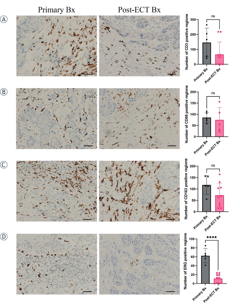
Figure 3.

Immunohistologically stained sections for lymphocytes CD3 (A), macrophages CD68 PMG1 (B) and CD163 (C), and blood vessels ERG (D), from primary biopsy (Primary Bx) and post-electrochemotherapy biopsy (post-ECT Bx) samples. Scale bar represents 50 μm. The number of positively stained regions ± standard error of the mean (SEM) is presented.
