
Figure 6. C. albicans-primed polyclonal T cells exacerbate ethanol-induced liver disease.
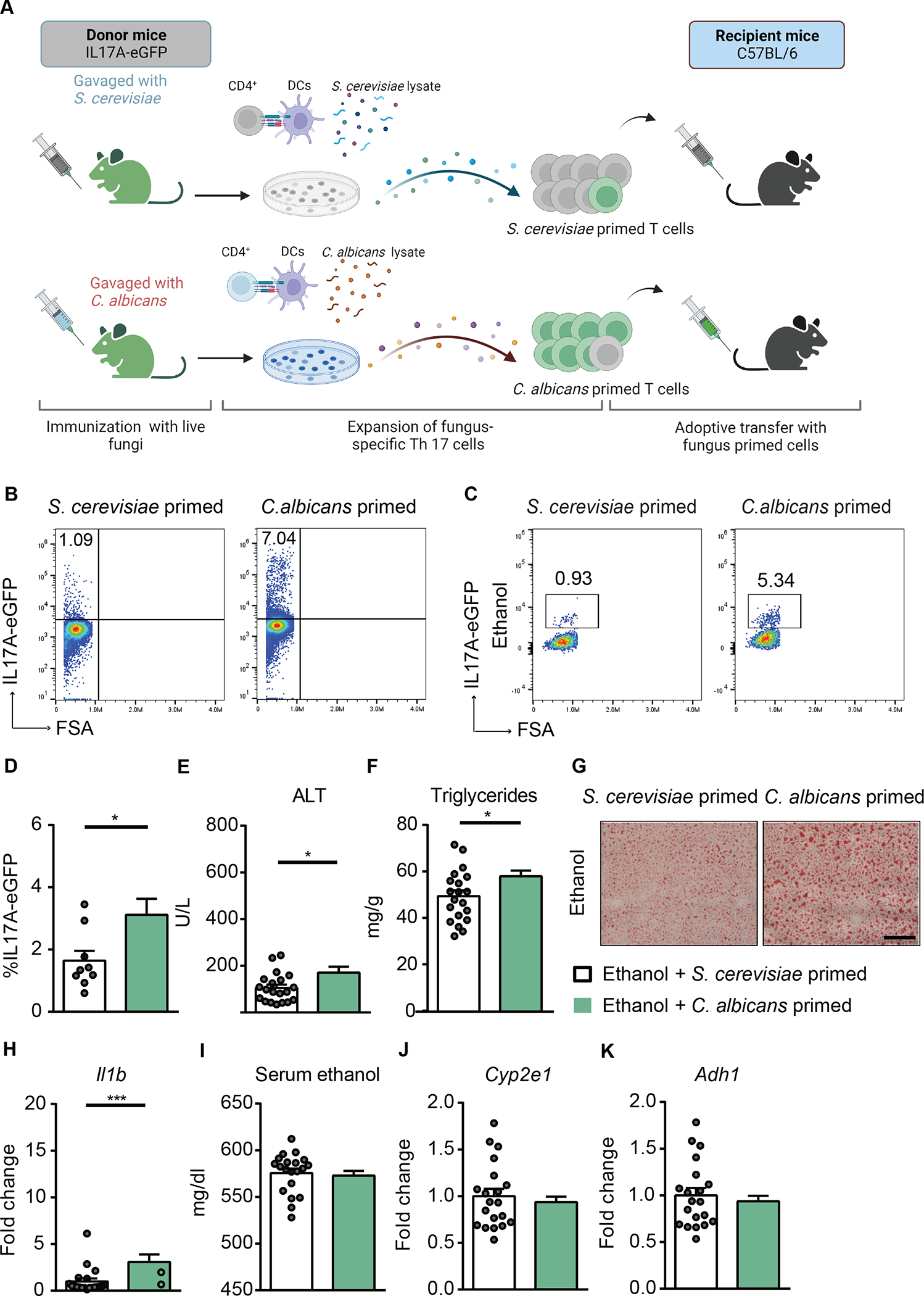
(A) Diagram of adoptive transfer of C. albicans-specific polyclonal T cells to mice. On day 0, IL17A-eGFP donor mice were gavaged with C. albicans or S. cerevisiae. On day 10, polyclonal CD4+ T cells from mesenteric lymph nodes and spleen of donor mice were co-cultured with bone marrow-derived dendritic cells in the presence of fungal lysates for 6 days. On day 16, polyclonal CD4+ T cells were injected intravenously to C57BL/6 mice (day 13 of chronic plus binge ethanol feeding). Created with BioRender.com. (B) The proportion of IL17A-eGFP+ cells among total CD4+ T cells after ex vivo stimulation for adoptive-transfer was assessed by flow cytometry. (C and D) IL17A-eGFP+ cells were detected in livers of ethanol-fed recipient mice at the time of collection. (E) Serum levels of ALT. (F) Hepatic triglyceride content. (G) Representative oil red O-stained liver sections (scale bar, 100 μm). (H) Hepatic levels of Il1b mRNA. (I) Serum levels of ethanol. (J and K) Hepatic levels of Cyp2e1 and Adh1 mRNAs. Figure 6 was conducted in 3 independent experiments. Results are expressed as mean±SEM. P values determined by 2-sided Student t test. *P<0.05, ***P<0.001. See also Figure S5.