
Figure 2-. Genome-wide association study of American black bear coat color.
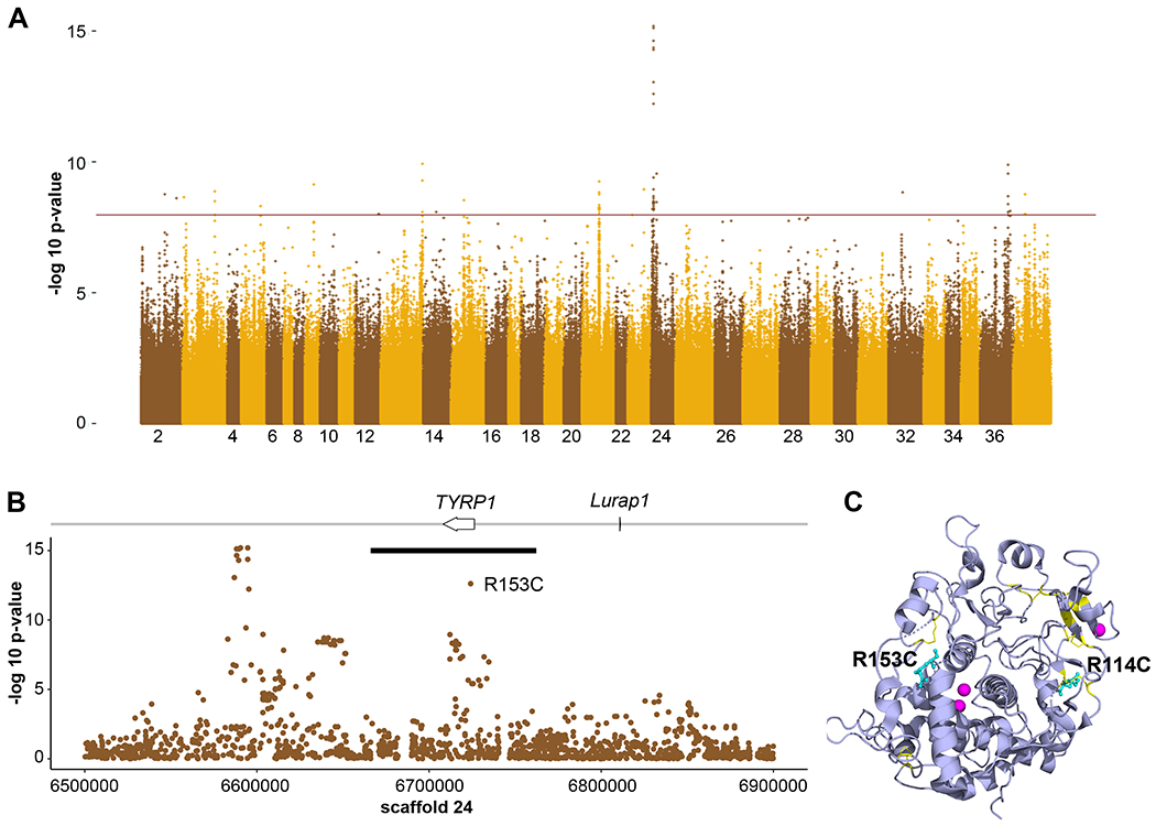
GWAS of both high and low coverage WGS data from Ursus americanus (n = 151) to identify loci associated with coat color. (A) A genome-wide Manhattan plot with a significance cut-off of 10−8 (horizontal brown line) identified a single strong peak on scaffold 24 (additional peaks in Table S6). (B) A detailed view of scaffold 24 surrounding the peak identified two genes, including TYRP1. The black bar denotes the length of the haplotype identified within the Nevada population that contains the R153C derived allele. (C) The locations of R114 and R153 (cyan, shown in atomic format) are shown within a ribbon diagram of the 3D structure of the human TYRP1 luminal domain (purple, from PDB ID 5M8L, 71). Cysteine residues involved in disulfide bonds are yellow and the zinc cofactors are magenta. Note the proximity of R114 to a native disulfide bond and of R153 to a break between two alpha helices that are part of the zinc binding region.