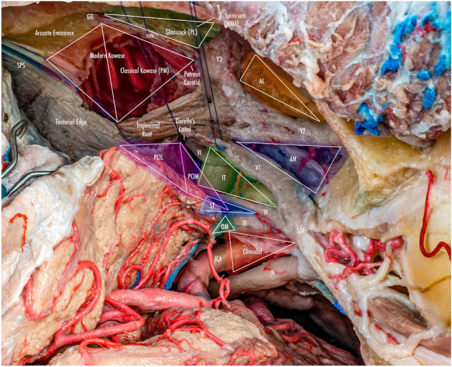
Fig. 1.

Anterolateral aspect of the middle cranial fossa depicting the triangles formed in this region. The roof and lateral aspect of the orbit have been drilled. The Sylvian fissure is shown splittled. The retractor is over the temporal lobe. From medial to lateral, the clinoidal triangle has been exposed after an anterior clinoidectomy has been done. It is between the optic and the oculomotor nerves and posteriorly bordered by the tentorial edge (Not shown). The oculomotor triangle (OM) is the site where the oculomotor nerve becomes extradural by entering the upper portion of the lateral wall of the cavernous sinus. Its margins are the anterior petroclinoial dural fold extending from the ACP to the petrous apex and the posterior petroclinoidal dural folds extending from the posterior clinoidal process to the petrous apex and medially by the interclinoidal dural fold. The supratrochlear triangle (ST), the space between the oculomotor and the trochlear nerves, has a posterior border drawn by a line at the dural entry point of these two nerves. The infratrochlear triangle (IT/Parkinson’s triangle) is lateral to the oculomotor and medial to the trochlear nerve. Its posterior border is the tentorial edge between these two nerves. The anteromedial triangle’s (AM/Mullan’s triangle) boundaries are the ophthalmic division of the trigeminal nerve medially and the maxillary division laterally. Its base is formed by a line connecting the superior orbital fissure to the foramen rotundum over the bony middle cranial fossa wall. The anterolateral triangle (AT) is formed medially by the maxillary division of the trigeminal nerve and laterally by the mandibular division (V3). The base is formed by a line connecting the foramen rotundum and the foramen ovale. Posteriorly over the middle cranial fossa, the Posteromedial and the posterolateral triangles can be found. The first of these two, the Posteromedial Middle Fosa Triangle (AKA Kawase’s triangle), is bordered laterally by the medial margin of the greater superficial petrosal nerve (GSPN). The petrous ridge is found medially. Anteriorly its boundary is the mandibular division of the trigeminus and laterally by V3. Posteriorly, the limit is the arcuate eminence. The posterolateral middle fossa triangle (Glasscock) is located laterally to the line where the GSPN crosses under V3 and the foramen spinosum. Its lateral border is a line between the foramen spinosum and the geniculate ganglion. Its base is GSPN. The paraclival triangles are the Inferomedial and Inferolateral triangles (PCIM and PCIL). The inferomedial triangle contains the dura forming the posterior wall of the cavernous sinus. It is delimited medially by a line extending from the posterior clinoid process to the dural entry of the abducens nerve. Its lateral border is a line extending from the posterior clinoid process to the dural entry of the trochlear nerve. Its base is the line extending from the dural entry of the abducens nerve and the trochlear nerve. Over the posterior surface of the clivus and the temporal bone, we can find the Inferolateral triangle (PCIL). Its anterior border is a line extending from the dural entry of the abducens nerve and the trochlear nerve's dural entry. Its lateral border is a line extending from the entrance of the trochlear nerve and the petrosal vein. Its posterior border is a line extending from the dural entry of the abducens nerve to the petrosal vein
